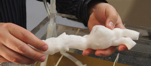
Because aortic aneurysms develop through an intricate interplay of environmental and genetic factors, they have been difficult to study both clinically and experimentally. The use of 3D printing to model aortic aneurysms at the University of Rochester represents a breakthrough for researchers seeking to study the disease. Speaking with sister publication MPMN, Ankur Chandra, associate professor of surgery and biomedical engineering at the university and a practicing vascular surgeon, explained the process.
April 14, 2014
“We start by creating a patient-specific 3-D model of an aneurysm. Next, we print it using a biologically accurate material that simulates the properties of the aortic wall, and then we mount it on a hemodynamic simulator, subjecting it to such tightly controlled hemodynamic parameters as intraluminal pressures, blood flows, and cardiac outputs,” Chandra told MPMN. As different hemodynamic and material properties change and stable aneurysms begin to fail, the researchers can study aneurysm behavior.
3D printing technology will enable many aortic devices to be tested in an ex vivo environment, Chandra added, allowing medical researchers to observe how the devices behave.
Read "New Dimension to 3D Printing: Modeling Aneurysms" for more about this technology.
Modeling aneurysms |
|
About the Author(s)
You May Also Like


